


INTRODUCTION
As the society is older aged, the number of patients of degenerative spine disease such as multilevel spinal stenosis is increased. Because the spine instrumentation surgery with wide decompression is effective treatment for degenerative spine disease, the number of spine surgery is increased, continuously [1].
Nevertheless, there are some complications after posterior screw fixation such as neurological or vascular injury, loss of curve correction, intraoperative pedicle fracture or loosening, dura injury, deep infection, and pseudarthrosis [2].
Because the old aged patients have high prevalence of osteoporosis, malnutrition, and often use long-term steroid, they are high-risk group of any fracture. But, the postoperative sacral fracture, distal part of lumbosacral arthrodesis, is rare and its pain is sometimes masked with postoperative pain, and that makes difficult early detection of sacral complication.
In a case of sacral fracture of distal level of fixed vertebral body after lumbosacral arthrodesis in old aged, we suggest the diagnosis and treatment of the fracture.
CASE REPORT
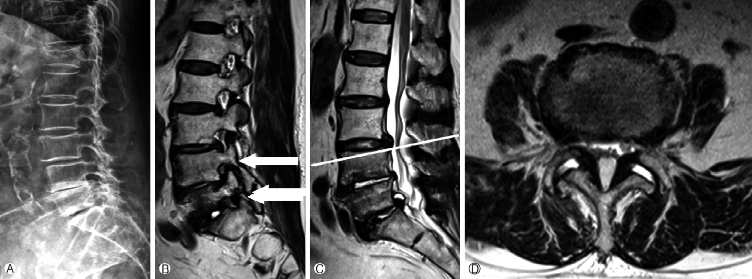
An 83-year-old female patient, previously operated L5/S1 level discectomy for leg pain. After 1-year previous operation, the patient complained lower back pain and recurred radiating pain on both legs, and did not be relieved although medical pain management and physical therapy programs. After lying down for 2-month at local clinic, she revisited our hospital for treatment. The plain radiographs and magnetic resonance images show spinal stenosis for multiple level, and spondylolisthesis of L3-4 level (Fig. 1). There is an evidence of osteopenia, on Bone DEXA scan. We operated her transforaminal lumbar interbody fusion for L4-5-S1 level, L3 laminectomy, posterior screw fixation for L3-4-5-S1 levels. There is no complication, intraoperative and post-operative.
After spine surgery, she felt the pain relieved, so started ambulation and rehabilitation therapy. But 1-month after ambulation, moderate low back and buttock pain occurred. The X-ray and CT images showed ŌĆśUŌĆÖ sacral fracture, classified Roy-Camille type 2 anterior displacement (Fig. 2). We suggested bed rest for 1-month for treatment of sacral fracture, and medical treatment of osteopenia and malnutrition state, so the pain is reduced and there is no evidence of further displacement of fracture.
So, the patient discharged, we check the serial X-ray examination for L-S spine in follow-up visit clinic, there is no evidence of aggravation (Fig. 3).
DISCUSSION
The lumbosacral arthrodesis is an effective way to manage spine degenerative disorders and deformities, vertebral fractures, and metastatic diseases [3-7]. However, in the patient having low bone density, such as osteopenia, osteoporosis, it is occurred failure of fusion, pullout of pedicle screw, even the fracture of pedicle screws or vertebral body of proximal or distal part of fixed vertebrae are uncommon [8-10]. In this case, the patient`s BMD was osteopenia but her bone quality was very poor. For 2-month before surgery and a week after spine surgery, the patient got a bed rest. So, sarcopenia state thought to be a one of the risk factors for sacral fracture. Besides, ambulation and rehabilitation are conducted carefully, sacral fracture was occurred.
Considering the reason that the patient had sacral fracture, we thought some hypothesis. Because of poor oral intake after surgery, the insufficient nutrition has influence of low bone density and probability of bone fusion. Malnutrition patient has more postoperative complications include reoperation [9]. Preoperative serum albumin levels have been an established marker for overall nutrition, and hypoalbuminemia measured by an albumin level less than 3.5 g/dL can be used as a proxy for malnourishment [11]. The patient`s initial albumin level was 2.8 g/dL and weight loss was 6 kg during 2-month (10% of total body weight). We recommend nutrition supply, strengthening muscles and maintaining weight through exercise before surgery. This theory is supported by published studies showing an independent association between recent weight loss resulting in malnutrition and an increased risk for postoperative complications [12].
Obesity, multi-segmental lumbosacral fusion, mismatched lumbar lordosis (LL)-pelvic incidence (PI) and high PI are also risk-factors of postoperative sacral fracture [13]. There was no PI to LL mismatch, postoperatively.
There are various treatments have been reported in the literature. These include conservative management, initial conservative treatment followed by surgical treatment after failure of conservative treatment, and primary surgical treatment. But there is no consensus on optimal treatment for sacral fracture after lumbosacral fusion. Three out of four cases reported by Vavken et al. were treated conservatively with good result [14]. In a radiographic study conducted by Wilde et al., 11 of 23 fractures were treated nonoperatively, and the other 12 went on to surgery with transiliac fixation [15]. Because there is similar improvement rate between surgical and conservative treatment [16]. The patient was tolerable for immobilization and pain. There was no neurological deficit and significant displacement. So, considering the re-operation risk of the patient we make a treatment plan conservative treatment by medical treatment of osteopenia and malnutrition state. And bed side physical treatment worked together by rehabilitation team.
As masked with postoperative pain, it is difficult to diagnose postoperative sacral fracture, especially around the operation site. Therefore, it should be performed CT or MR images when patients complain of renewed lower back pain or buttock pain.
After occurring sacral fracture, we checked the serial simple radiograph, CT scan and estimated patient symptom. In the present patient did not opted for additional surgery because pain and disability were mild. Most importantly, if the risk factors are corrected before operation, we predict the probability of fracture decreased.
CONCLUSION
After spine surgery, sacral fracture of distal fusion part is not common. To diagnosis sacral fracture, the physician should pay attention to complain of pain. If the sacral fracture is dia gnosed, the physician can choose surgical treatment or medical conservative management. Such as this patient, old-aged, having lots of underlying disease, we suggest medical conservative management instead of surgery. And it may be accompanied the sufficient nutrition, treatment of osteopenia and physical therapy.